|
||||
|
|
BONUS CONTENT FROM NURSING MADE INCREDIBLY EASY! Cholecystectomy:
Take a look at two options About 700,000 cholecystectomies are performed each year for patients diagnosed with gallbladder disease, making it one of the most routine surgeries performed. The minimally invasive laparoscopic cholecystectomy is the standard of care for most patients needing cholecystectomy. Open surgery is an option for patients who can’t have laparoscopic surgery. In this article, I’ll review
gallbladder pathophysiology and your role in caring for a patient
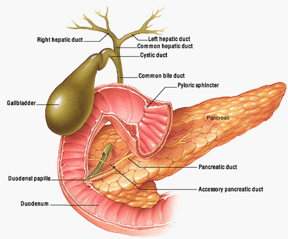
who needs a cholecystectomy. For details on gallbladder anatomy,
see the image to the right. Gallbladder dysfunction Gallstones are caused by changes in the composition of bile, especially bile salts, phospholipids, bilirubin, and cholesterol. When these solids are supersaturated in the gallbladder, gallstones may form. The gallbladder secretes mucus and proteins that promote cholesterol crystal formation, which is the precursor for stone formation in supersaturated bile. Impaired gallbladder motility, biliary stasis, and changes in bile content can lead to stone formation. For details, see Not the rolling stones. The risk of gallstones increases with advancing age, and women face a higher risk than men. Other risk factors include white race, obesity, sedentary lifestyle, alcoholism, pregnancy, rapid weight loss, oral contraceptive use, high-fat diet, diseases of the ileum, terminal ileum resection, parenteral nutrition, dyslipidemia, use of cholesterol-lowering drugs, cirrhosis, hereditary spherocytosis, and hemolytic anemia. Diagnosing gallbladder trouble To evaluate a patient for possible cholecystitis, obtain specimens for lab tests, including a complete blood cell (CBC) count, liver function tests, serum amylase and lipase levels, and pregnancy test. In a patient with cholecystitis, the CBC count typically shows elevated white blood cell count related to inflammation; aspartate aminotransferase, alanine aminotransferase, and alkaline phosphate levels may also be elevated in common bile duct obstructions. The healthcare provider will order imaging studies, such as an ultrasound of the right upper quadrant (the standard imaging test). This can reveal gallstones, gallbladder wall hickening, and pericholecystic fluid. In cases where an ultrasound won’t yield clear images (for example, if the patient is obese), the healthcare provider may order a hydroxyiminodiacetic acid (HIDA) scan. This study can help confirm cholecystitis by demonstrating abnormal gallbladder function. A pear-shaped storage tank Treatment options Laparoscopic cholecystectomy has many benefits for patients, including a shorter hospital stay, less pain and scarring, less trauma to tissues, a shorter healing and recovery time, and a quick return to normal activities (usually within 3 to 5 days). Instead of making a 5- to 7-inch-long (12.5- to 17.5-cm-long) abdominal incision, the surgeon makes just four small stab wounds (see One big or four small?). He inserts trocars at all incision sites to provide ports of entry. This minimally invasive surgery requires a special arrangement of equipment to provide maximum visualization. The surgeon insufflates carbon dioxide into the abdominal cavity through the Verres needle to establish pneumoperitoneum. Pneumoperitoneum facilitates visualization of abdominal structures and instrument manipulation. The surgeon identifies the cystic duct and artery and looks for stones in the biliary tree with the laparoscopic instruments. After dividing the cystic duct and artery, he dissects the gallbladder away from the liver using a laparoscopic instrument connected to cautery for hemostasis. After freeing the gallbladder, he removes it through the umbilical incision. He then checks the liver bed for bleeding and the abdomen for bile and stones. The peritoneal cavity is decompressed of carbon dioxide and all incisions are closed. The gallbladder and its contents are sent to pathology for analysis. At any time during a laparoscopic procedure, the surgeon may convert to an open procedure if complications arise that threaten patient safety. Possible problems requiring open surgery include adhesions that impair the surgeon's ability to visualize abdominal structures, an injury to the bile duct or associated organs, gallbladder edema, and bleeding. Open gallbladder surgery is much like the laparoscopic version. The surgeon examines the biliary tree and cystic duct and handles them in the same surgical manner. But in the open procedure, the surgeon can use his hands to palpate for stones and can directly examine the gallbladder before removal. Because a patient who needs open surgery may have major medical issues, the fragile tissues of the inflamed gallbladder put him at greater risk for bleeding or bile spillage. (Bile spillage also can occur during the laparoscopic procedure because of gallbladder inflammation or perforation with a laparoscopic instrument.) If bile is spilled, the surgeon irrigates the abdomen with 0.9% sodium chloride solution to prevent peritonitis. He also may place a drain in the subhepatic space. After separating the gallbladder from the liver bed and sending it to pathology, the surgeon closes the muscle layers of the incision with durable sutures that will withstand abdominal pressure. He may approximate the skin incision line with sutures or staples, depending on his preference. Before the procedure Prepare a patient undergoing laparoscopic surgery for postoperative shoulder and neck pain secondary to phrenic nerve irritation from the carbon dioxide used to insufflate the peritoneum. This minor discomfort may last a few days, but may be relieved by changing position. For patients having an open cholecystectomy, review the importance of incentive spirometry, deep breathing, and coughing after surgery to reduce the risk of atelectasis and pneumonia. Also review the importance of early and aggressive ambulation to help reduce the risk of venous thromboembolism (VTE). On the day of surgery, verify patient identification
and review the patient’s medical history and physical, surgical
history (including a personal or family history of anesthesia
problems), lab results, limitations for positioning the patient
on the OR table, and allergies. Perform medication reconciliation,
confirm her N.P.O. status, confirm evidence of the informed consent
process, and discuss postoperative care and pain management, including
how to use a patient-controlled analgesia (PCA) pump, if applicable. After the procedure Postoperative care for patients is the same for both types of surgery. Regularly assess your patient’s level of consciousness and vital signs and monitor her closely for signs and symptoms of bleeding. Use a valid and reliable pain intensity rating scale to assess her pain and provide optimal pain management. If she has a PCA pump, review how to use it. Check dressings for drainage and incision sites for signs of infection. Persistent pain unrelieved by analgesics, persistent fever over 101° F (38° C), chills, abdominal distension, anorexia, persistent nausea and vomiting, and jaundice may indicate bile duct injury and should be reported immediately to the surgeon. Place a patient who’s had an open cholecystectomy in low Fowler’s position. When she’s alert, encourage her to use the incentive spirometer. Show her how to splint her incision when necessary. After laparoscopic surgery, position the patient in a left side-lying Sims position to move retained pockets of carbon dioxide away from the diaphragm and decrease discomfort. Encourage early and aggressive ambulation after surgery to help prevent VTE. Evaluate the patient’s hemoglobin and hematocrit levels and notify the healthcare provider if they’re abnormal. Follow the American College of Chest Physicians evidence-based clinical practice guidelines for VTE prophylaxis, depending on level of thromboembolism risk. The patient should start with clear liquids and gradually advance her diet as tolerated. She should eat a high-fiber diet and drink plenty of fluids unless contraindicated. If she has cramping in the right upper abdominal quadrant, advise her to reduce her fat intake. Explain that feces will pass through the colon faster after cholecystectomy. She may need a bile acid binder (such as cholestyramine or colestipol) if she develops chronic diarrhea. Before discharge, teach your patient to call her healthcare provider if she has excessive or abnormal bleeding, a fever greater than 101° F, jaundice, abdominal distension or pain, persistent cough, or shortness of breath. Teach her about her prescribed pain medications and how to monitor wound healing. Also teach her to monitor her bowel habits, especially if she’s been prescribed a bile acid binder. Tell her that bile acid binders can cause constipation and heartburn and can interact with other drugs, including beta-blockers, thiazide diuretics, and warfarin. She should take medications at least 1 hour before or 4 to 6 hours after taking the bile acid binder and should call her healthcare provider immediately if she has any noticeable physical changes. Staying well
References Source: Nursing2009. February 2009. |
|||